The Physics of Imaging the Human Body
Pre/Post Test |
Name __________________________________ |
| |
|
|
|
| |
1. |
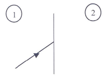
A periodic sound wave is travelling in a body. It enters a
region with another type of tissue. The densities of the first and second
tissue types are essentially the same but the bulk modulus of the second tissue
is smaller than that of the first tissue. The figure below shows the direction
of travel for the sound wave. |
| |
 |
| |
|
|
|
| (2) |
a. |
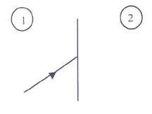
Describe what happens to the wave when it hits the interface between the two tissues. You can draw on the figure below to help with your description. |
| |
 |
| |
|
|
|
| (2) |
b. |
Compare the sound speeds in the two tissues. Explain how you made your choice.
|
| |
|
|
|
| (2) |
c. |
What happens to the wavelength of the sound when it enters tissue 2? Explain.
|
| |
2. |
The table gives some physical data for 2 types of tissue: |
| |
|
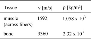
Data from Diagnostic Ultrasound, Matthew Hussey, Blackie & Son Limited (London, 1975). |
| |
|
|
|
| (2) |
a. |
How do the acoustic impedances compare between the two tissue types?
|
| (2) |
b. |
If a sound wave in muscle hits bone, would a significant portion of the wave's energy be transmitted into the bone? Explain how you know.
|
| (2) |
3. |
An ultrasound pulse is introduced into a body. Reflected pulses from within the body are received by the transducer and displayed on a CRT. An idealized display is shown to the right. This is what an A-scan ultrasound instrument displays. The horizontal axis on the display shows time. Each division represents 0.005 milliseconds. |
 |
| |
|
|
|
| |
Assume that the speed of sound in all the tissues encountered is 1550 [m/s].
How far is it to the farthest tissue interface seen by this instrument? Explain.
|
| (2) |
4. |
Describe how a B-scan ultrasound instrument can
produce a two-dimensional image of an internal section of a body.
|
| (2) |
5. |
A sound wave in muscle is incident onto bone. Its incident angle is 20°, as shown below. What would be the refracted angle of the sound wave in the bone?
You can use the data from problem 2, if desired. Show all work.
 |
| |
6. |
An ultrasound pulse is incident on moving blood, as shown to the right. The transducer can detect the pulse reflected from blood. |
 |
| (2) |
a. |
How will the frequency of the reflected pulse compare with the
orginal pulse? Explain how you know.
|
| |
|
Now the incident pulse is oriented as shown to the right. |
 |
| (2) |
b. |
How will the frequency of the reflected pulse compare to that of
the original pulse?
|
| |
|
|
|
| |
|
|
|
| |
|
|
|
| |
|
|
|

